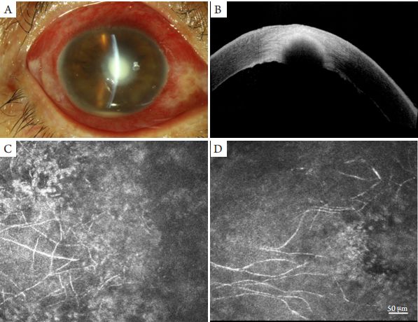
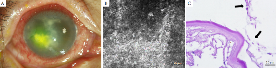
Retrocorneal fungal infection near the corneal limbus (A), slit lamp examination showing a deep stroma lesion with intact epithelium (B),and confocal microscopy images showing the hyphae in deep stroma (C), and the hyphae in the endothelium layer (D).
图2 典型角膜后部真菌感染患者裂隙灯及共聚焦图像
Figure 2 Representitive images of slit lamp and confocal microscope of retrocorneal fungal infection
Retrocorneal fungal infection near the corneal limbus (A), slit lamp examination showing a deep stroma lesion with intact epithelium (B),and confocal microscopy images showing the hyphae in deep stroma (C), and the hyphae in the endothelium layer (D).
图3 典型角膜后部真菌感染患者的激光共聚焦显微镜检查图像
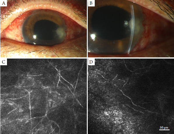
Figure 3 Representitive images of confocal microscope of retrocorneal fungal infection
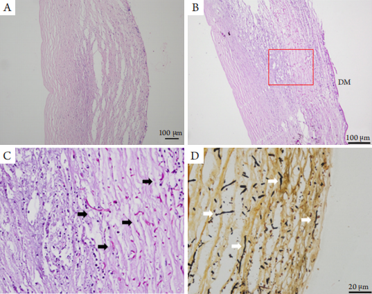
(A)Retrocorneal fungal infection with inflammation cells infiltration and storma necrosis, PAS staining shown hyphae in deep stroma and near the DM (B), black arrows shown stained hyphae (C), and white arrows shown black and brown stained hyphae with GMS staining (D).
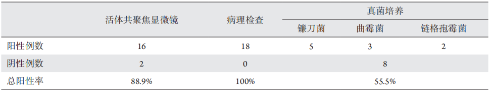
表1 角膜后部真菌感染患者活体共聚焦显微镜、病理检查和真菌培养诊断阳性率比较
Table 1 Comparison of positive rates of in vivo confocal microscopy, pathological examination and fungal culture in retrocorneal fungal infection
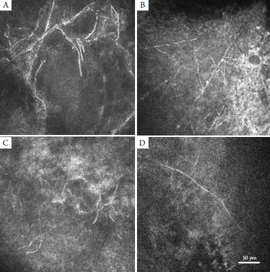
图5 角膜深层真菌感染患者的裂隙灯、共聚焦及病理PAS图像
Figure 5 Representitive images of slit lamp, confocal microscope and PAS stain
(A) Retrocorneal lesion with hypopyon, no hyphae in deep stroma with confocal microscopy examination (B), and black arrows shown the hyphae on the back of DM with PAS staining (C).
1. 山东省自然科学基金 (ZR2015YL026);青岛市市南区科技发展资金项目 (2018-4-030-YY)。 This work was
supported by the Natural Science Foundation of Shandong (ZR2015YL026) and the Science and Technology Program of Qingdao South District (2018-4-030-
YY), China.
参考文献
1. Xie L, Zhong W, Shi W, et al. Spectrum of fungal keratitis in north
China[ J]. Ophthalmology, 2006, 113(11): 1943-1948.
2. Erie JC,Mc Laren JW, Pate l SV.Confocal microscopy in
ophthalmology[ J]. Am J Ophthalmol, 2009, 148(5): 639-646.
3. Niederer RL, McGhee CN. Clinical in vivo confocal microscopy of
the human cornea in health and disease[ J]. Prog Retin Eye Res, 2010,
29(1): 30-58.
4. 刘瑞, 李冰, 徐鼎, 等. 活体共聚焦显微镜观察视网膜激光光凝术
对角膜上皮下神经的影响[ J]. 眼科学报, 2018, 33(2): 101-107.
LIU R, LI B, XU D, et al. Effect of retinal photocoagulation on
corneal sub-basal nerve observed by in vivo confocal microscopy[ J].
Yan Ke Xue Bao, 2018, 33(2): 101-107.
5. Villani E, Baudouin C, Efron N, et al. In vivo confocal microscopy of
the ocular surface: from bench to bedside[ J]. Curr Eye Res, 2014,
39(3): 213-231.
6. 王楠, 赵桂秋, 高昂, 等. 刮片镜检法联合激光扫描共焦显微镜快
速诊断真菌性角膜炎的临床评价[ J]. 中华实验眼科杂志, 2013,
31(5): 493-495.
WANG N, ZHAO GQ, GAO A, et al. Clinical evaluation
of rapid diagnosis of fungal keratitis by the combination of corneal
scraping with laser scanning confocal microscopy[ J]. Chinese Journal
of Experimental Ophthalmology, 2013, 31(5): 493-495.
8. Thomas PA, Kaliamurthy J. Mycotic keratitis: epidemiology, diagnosis
and management[ J]. Clin Microbiol Infect, 2013, 19(3): 210-220.
9. Chidambaram JD, Prajna NV, Palepu S, et al. In vivo confocal
microscopy cellular features of host and organism in bacterial,
fungal, and acanthamoeba keratitis[ J]. Am J Ophthalmol, 2018,
190(6): 24-33.
10. 史伟云, 牛晓光, 王富华, 等. 真菌性角膜炎药物治疗后转归的共
焦显微镜观察[ J]. 中华眼科杂志, 2005, 41(7): 614-619.
SHI WY, NIU XG, WANG FH, et al. Evaluation of
antifungal chemotherapeutic effects on fungal keratitis by confocal
microscopy[ J]. Chinese Journal of Ophthalmology, 2005, 41(7):
614-619.
11. 程钧, 翟华蕾, 王君怡, 等. 角膜后部真菌感染的临床特点和治疗
策略[ J]. 中华眼科杂志, 2017, 53(10): 758-765.
CHENG J, ZHAI HL, WANG JY, et al. Clinical features and
treatments of retrocorneal fungal infection[ J]. Chinese Journal of
Ophthalmology, 2017, 53(10): 758-765.
12. 梁庆丰, 孙旭光, Antoine L. 活体共聚焦显微镜在感染性角膜炎
诊治中的应用[ J]. 中华眼科杂志, 2013, 49(10): 951-955.
LIANG QF, SUN XG, LABBE Antoine. Role of in vivo
confocal microscopy in the management of infectious keratitis[ J].
Chinese Journal of Ophthalmology, 2013, 49(10): 951-955.
13. Kanavi MR, Javadi M, Yazdani S, et al. Sensitivity and specificity of
confocal scan in the diagnosis of infectious keratitis[ J]. Cornea, 2007,
26(7): 782-786.
14. Chidambaram JD, Prajna NV, Larke NL, et al. Prospective study
of the diagnostic accuracy of the in vivo laser scanning confocal
microscope for severe microbial keratitis[ J]. Ophthalmology, 2016,
123(11): 2285-2293.
15. 高欢欢, 张平, 林健贤, 等. 三种特殊染色方法在角结膜原位癌基
底膜染色中的应用[ J]. 眼科学报, 2018, 33(1): 32-36.
GAO HH, ZHANG P, LIN JX, et al. Application of
three special staining methods in the basement membranes in corneal
and conjunctival carcinoma in situ[ J]. Eye Science, 2018, 33(1): 32-36.
16. Kheirkhah A, Syed ZA, Satitpitakul V, et al. Sensitivity and specificity
of laser-scanning in vivo confocal microscopy for filamentous fungal
keratitis: role of observer experience[ J]. Am J Ophthalmol, 2017,
179(7): 81-89.
17. Winchester K, Mathers WD, Sutphin JE. Diagnosis of Aspergillus
keratitis in vivo with confocal microscopy[ J]. Cornea, 1997, 16(1):
27-31.
18. Chidambaram JD, Prajna NV, Larke N, et al. In vivo confocal
microscopy appearance of Fusarium and Aspergillus species in fungal
keratitis[ J]. Br J Ophthalmol, 2017, 101(8): 1119-1123.
'%20fill='white'%20fill-opacity='0.01'/%3e%3cmask%20id='mask0_3477_29692'%20style='mask-type:luminance'%20maskUnits='userSpaceOnUse'%20x='0'%20y='0'%20width='16'%20height='16'%3e%3crect%20id='&%23232;&%23146;&%23153;&%23231;&%23137;&%23136;_2'%20x='16'%20width='16'%20height='16'%20transform='rotate(90%2016%200)'%20fill='white'/%3e%3c/mask%3e%3cg%20mask='url(%23mask0_3477_29692)'%3e%3cpath%20id='&%23232;&%23183;&%23175;&%23229;&%23190;&%23132;'%20d='M14%205L8%2011L2%205'%20stroke='%23333333'%20stroke-width='1.5'%20stroke-linecap='round'%20stroke-linejoin='round'/%3e%3c/g%3e%3c/g%3e%3c/svg%3e)