Notes: (A) Before the laser treatment: pigmented nevus located at the upper and lower eyelid margins of the left eye, adjacent to the lacrimal punctum and extending to the eyelid conjunctiva. (B) One month after the seventh laser treatment: evidence of partial sparse eyelashes, absence of lesion recurrence, no scar hyperplasia, and a smooth eyelid margin without signs of entropion or ectropion. (C) Pathological diagnosis: pigmented nevus OS. Pathological findings: scattered nevus cells beneath the stratified squamous epithelium, with preserved cellular polarity and no discernible cellular atypia. (HE staining, ×200)
见图2
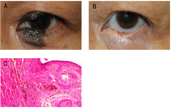
图2 患者男,25岁,左眼下睑色素痣激光治疗前后眼外观对比图以及激光切除肿物病理检查图
Figure 2 Pigmented nevus (OS) before and after laser treatment and pathological examination in a 25-year-old male
Notes: (A) Before the laser treatment: a black lesion grew along the eyelid margin on the lateral side of the lower eyelid, about 12 mm×5 mm in size, with a clear boundary, involving the root of eyelashes and conjunctiva. (B) One month after the fourth laser treatment: absence of lesion recurrence, no scar hyperplasia, a smooth eyelid margin without signs of entropion or ectropion, and partial sparse eyelashes in the treatment area. (C) Pathological diagnosis: pigmented nevus OS. Pathological findings: scattered nevus cells beneath the stratified squamous epithelium, with preserved cellular polarity and no discernible cellular atypia. (HE staining, ×200)
见图3
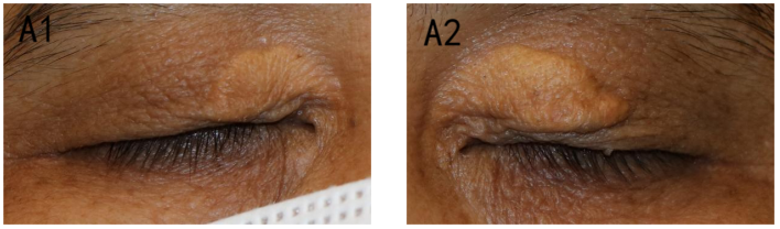
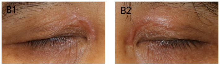
图3 患者女,48岁,双眼上睑黄色瘤激光治疗前后眼外观对比图
Figue 3 Xanthelasma (OU) before and after laser treatment in a 48-year-old female
Notes: (A1 & A2) Before the laser treatment: there were flat yellow lesions on the inner side of the upper eyelid of both eyes with clear edges. The size of the lesions in the right eye was about 10 mm× 6 mm, and that in the left eye was about 15 mm× 6mm. (B1 & B2) One month after the fifth laser treatment: there was no lesion recurrence, minimal scar hyperplasia and pigmentation.
见图4
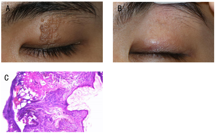
图4 患者男,18岁,左眼上睑疣激光治疗前后眼外观对比图以及激光切除肿物病理检查图
Figure 4 Eyelid molluscum (OS) before and after laser treatment and pathological examination in an 18-year-old male
(A) Before the laser treatment: a dense granular lesion extending vertically from the eyelid margin to the eyebrow root, ranging about 8 mm×20 mm. (B) One month after the third laser treatment: no evidence of lesion recurrence, scar hyperplasia, or trichiasis. (C) Pathological diagnosis: eyelid molluscum OS. Pathological findings: proliferative and degenerative changes in the stratified squamous epithelium. The epithelium showed incomplete keratinization alongside hyperkeratosis. (HE staining, ×200)
见图5
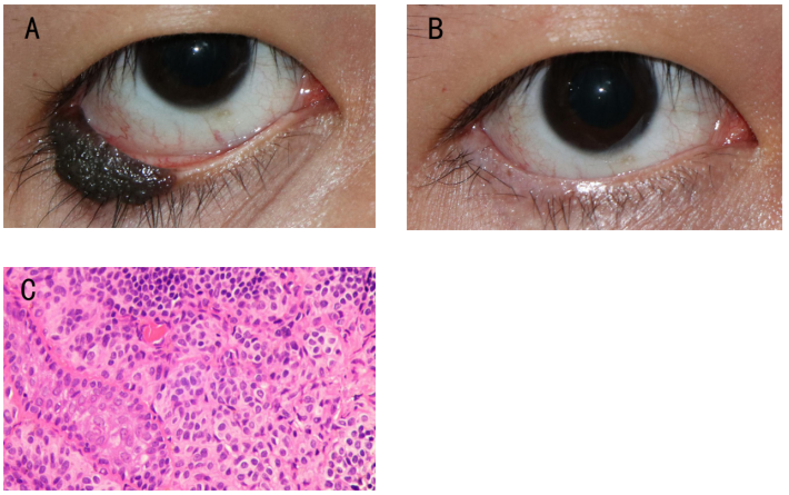
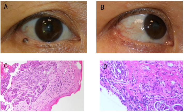
图5 患者女,48岁,右眼下睑肿物激光治疗前后眼外观对比图以及激光切除肿物病理检查图
Figure 5 Malignant melanoma (OD) of the eyelid before and after laser treatment and pathological examination in a 48-year-old female
(A) Before the laser treatment: black lesion located at the root of the eyelashes of the lower eyelid, about 2 mm×2 mm in size, and not involved gray line. (B) One-month post-laser treatment: no recurrence of the lesion, no evidence of scar hyperplasia, no signs of entropion or ectropion, and partial sparse eyelashes in the treated area. (C) Pathological diagnosis (HE staining, ×200): malignant melanoma OD. (D) One-month after laser treatment: a controlled surgical excision of the lesion area was performed. Histological examination revealed fibrous tissue proliferation and degeneration, with minimal lymphocytic infiltration and no evident tumor tissue. (HE staining, ×200)
3. Goto H, Yamakawa N, Komatsu H, et al. Epidemiological characteristics of malignant eyelid tumors at a referral hospital in Japan[J]. Jpn J Ophthalmol, 2022, 66(4): 343-349.
4. Gniesmer S, Sonntag SR, Schiemenz C, et al. Diagnosis and treatment of malignant eyelid tumors[J]. Ophthalmologie, 2023, 120(3): 262-270.
5. Liu J, Sun J, Wang Z, et al. Treatment of divided eyelid nevus with orbicularis oculi myocutaneous flap: report of 17 cases[J]. Ann Plast Surg, 2020, 85(6): 626-630.
6. Torto FL, Losco L, Bernardini N, et al. Surgical treatment with locoregional flaps for the eyelid: a review[J]. Biomed Res Int, 2017, 2017: 6742537.
7. Zhao S, Duan J, Zhang J, et al. Evaluation of meibomian gland function after therapy of eyelid tumors at palpebral margin with super pulse CO2 laser[J]. Dis Markers, 2022, 2022: 8705436.
8. Zhang J, Duan J, Gong L. Super pulse CO2 laser therapy for benign eyelid tumors[J]. J Cosmet Dermatol, 2018, 17(2): 171-175.
9. Rentka A, Grygar J, Nemes Z, et al. Evaluation of carbon dioxide laser therapy for benign tumors of the eyelid margin[J]. Lasers Med Sci, 2017, 32(8): 1901-1907.
10. 富秋涛, 魏宁, 孟辉, 等. 超脉冲CO2激光治疗睑缘分裂痣疗效观察[J]. 激光生物学报, 2019, 28(1): 80-83.
Fu QT, Wei N, Meng H, et al. Efficacy of ultrapulse CO2 laser treatment divided nevus of the eyelid[J]. Acta Laser Biol Sin, 2019, 28(1): 80-83.
11. 曾颖, 罗益金, 占魁. CO2激光治疗睑缘色素痣的临床观察[J]. 应用激光, 2020, 40(4): 768-771.
Zeng Y, Luo YJ, Zhan K. Clinical observation on palpebral margin melanocytic naevus with super pulsed CO2 laser treatment[J]. Appl Laser, 2020, 40(4): 768-771.
12. Mao Z, Lin BY, Huang YD, et al. Microscopic treatment of benign eyelid margin lesions with ultrapulse carbon dioxide (CO2) laser[J]. J Cosmet Laser Ther, 2021, 23(7-8): 184-187.
13. Varde MA, Murali KV, Wiechens B. Surgical treatment of eyelid tumors[J]. HNO, 2018, 66(10): 743-750.
14. Then SY, Malhotra R. Superiorly hinged blepharoplasty flap for reconstruction of medial upper eyelid defects following excision of xanthelasma palpebrum[J]. Clin Exp Ophthalmol, 2008, 36(5): 410-414.
15. 王越, 李洋, 侯志嘉, 等. 眼睑分裂痣的新分类法及整复手术效果 [J] . 中华眼科杂志, 2022, 58(9) : 676-681.
Wang Y, Li Y, Hou ZJ, et al. A new classification method of eyelid divided nevi and the effect of plastic surgical treatment[J]. Chin J Ophthalmol, 2022, 58(9): 676-681.
16. Chin JKY, Yip W, Young A, et al. A six-year review of the latest oculoplastic surgical development[J]. Asia Pac J Ophthalmol, 2020, 9(5): 461-469.
17. Cho HJ, Lee W, Jeon MK, et al. Staged mosaic punching excision of a kissing nevus on the eyelid[J]. Aesthetic Plast Surg, 2019, 43(3): 652-657.
18. Al-Niaimi F. Ultrapulsed CO2 ablation in the treatment of xanthelasma palpebrarum: high satisfaction treatment with low recurrence[J]. J Dermatolog Treat, 2022, 33(2): 1116-1118.
19. Li D, Lin SB, Cheng B. CO2 laser treatment of xanthelasma palpebrarum in skin types III-IV: efficacy and complications after 9-month follow-up[J]. Photobiomodul Photomed Laser Surg, 2019, 37(4): 244-247.
20. Krupa Shankar D, Chakravarthi M, Shilpakar R. Carbon dioxide laser guidelines[J]. J Cutan Aesthet Surg, 2009, 2(2): 72-80.
21. van Gemert MJC, Bloemen PR, Wang WY, et al. Periocular CO2 laser resurfacing: severe ocular complications from multiple unintentional laser impacts on the protective metal eye shields[J]. Lasers Surg Med, 2018, 50(10): 980-986.
'%20fill='white'%20fill-opacity='0.01'/%3e%3cmask%20id='mask0_3477_29692'%20style='mask-type:luminance'%20maskUnits='userSpaceOnUse'%20x='0'%20y='0'%20width='16'%20height='16'%3e%3crect%20id='&%23232;&%23146;&%23153;&%23231;&%23137;&%23136;_2'%20x='16'%20width='16'%20height='16'%20transform='rotate(90%2016%200)'%20fill='white'/%3e%3c/mask%3e%3cg%20mask='url(%23mask0_3477_29692)'%3e%3cpath%20id='&%23232;&%23183;&%23175;&%23229;&%23190;&%23132;'%20d='M14%205L8%2011L2%205'%20stroke='%23333333'%20stroke-width='1.5'%20stroke-linecap='round'%20stroke-linejoin='round'/%3e%3c/g%3e%3c/g%3e%3c/svg%3e)