
Choroidal Analysis of Polypoidal Choroidal Vasculopathy by Spectral Domain Optical Coherence Tomography
阅读量:913
DOI:10.3969 / j.issn.1000-4432.2014.01.004
发布日期:2025-07-18
作者:
Chunyi Wei ,Ran Liu ,Jiaqing Li ,Yu Yang ,Xiaoyan Ding
展开更多 '%20fill='white'%20fill-opacity='0.01'/%3e%3cmask%20id='mask0_3477_29692'%20style='mask-type:luminance'%20maskUnits='userSpaceOnUse'%20x='0'%20y='0'%20width='16'%20height='16'%3e%3crect%20id='&%23232;&%23146;&%23153;&%23231;&%23137;&%23136;_2'%20x='16'%20width='16'%20height='16'%20transform='rotate(90%2016%200)'%20fill='white'/%3e%3c/mask%3e%3cg%20mask='url(%23mask0_3477_29692)'%3e%3cpath%20id='&%23232;&%23183;&%23175;&%23229;&%23190;&%23132;'%20d='M14%205L8%2011L2%205'%20stroke='%23333333'%20stroke-width='1.5'%20stroke-linecap='round'%20stroke-linejoin='round'/%3e%3c/g%3e%3c/g%3e%3c/svg%3e)
关键词
spectral domain optical coherence tomography
subfoveal choroidal thickness
maximal choroidal vessel diameter
polypoidal choroidal vasculopathy
摘要
Purpose: This study was designed to measure the changes in the subfoveal choroidal thickness (SFCT) and choroidal maximal vessel diameter (MVD) of the affected and unaffected fellow eyes in patients with polypoidal choroidal vasculopathy (PCV) and compare them to healthy controls.
Methods : In this cross-sectional observational clinical study, SFCT and MVD were measured in both eyes of 53 patients with unilateral PCV. PCV eyes were subgrouped into group A and unaffected fellow eyes into group B. All patients were diagnosed with PCV by fundus fluorescein angiography (FFA) and indocyanine green angiography (ICGA). Sixty age- and gender- matched healthy subjects were enrolled in the control group (group C).
Results: No statistical difference was observed among groups in age and gender. Overall, SFCT was correlated with MVD in all subjects (P<0.001; correlation coefficient: 0.759). P values were<0.001 with a correlation coefficient of 0.686, 0.801, and 0.808 in groups A, B, and C, respectively. No statistical significance was noted in SFCT among groups A (266.45±99.51 μm), B (269.57 ± 105.10 μm), and C (243.83±99.68 μm) (P=0.335). However, the MVD in group A was (202.55±72.45 μm), significantly larger than that in group C (166.45±56.18 μm,P=0.008), while the MVD in group B (194.75±85.27 μm) was equally significantly greater than that in group C (166.45±56.18 μm)(P=0.038).
Conclusion: For both PCV patients and healthy subjects, SFCT was positively correlated with MVD. No statistical sig nificance was noted in SFCT between PCV eyes and unaffected fellow/normal eyes. However, MVD was significantly larger in the PCV affected eyes than in unaffected fellow or normal control eyes, suggesting that MVD could be considered as a sensitive indicator to evaluate choroidal perfusion in PCV patients.
全文
The choroid is the primary nutritional source for all ocular tissues and plays a vital role in the metabolism of eyeballs. The choroidal structural or blood flow dynamic changes are probably associated with the incidence and progress of retinal diseases, such as polypoidal choroidal vasculopathy(PCV), etc.1. Choroid vascular pathological dilatations may underlie the pathogenesis of PCV2. Terasaki et al3. Obtained submacular tissue specimens from PCV patients in traoperatively and found dilated thin-walled vessels in Bruch’s membrane with multiple lesions in poly poidal or cluster shapes. Indocyanine green angiography revealed the formation of high-perfusion an giopathy of the choroidal vasculature and polypoidal structure, further confirming the signs of PCV. At present, this is considered the gold standard for PCV diagnosis.
The advances in optic coherence tomography (OCT) have led to a deeper understanding of the underlying pathogenesis of PCV. The emergence of new-generation spectral domain optical coherence tomography (SD-OCT) allows explicit observation of each layer structure of the retina and choroid4,5. In the present study, the alterations in choroidal vascu lature were observed in PCV patients by using SD-OCT to measure the subfoveal choroidal thickness (SFCT) and choroidal maximal vessel diameter (MVD) in affected and unaffected fellow eyes.
Materials and methods
Study subjects
The choroid is the primary nutritional source for all ocular tissues and plays a vital role in the metabolism of eyeballs.The choroidal structural or blood flow dynamic changes are probably associated with the incidence and progress of retinal diseases, such as polypoidalchoroidal vasculopathy (PCV), etc.1.
Choroid vascular pathological dilatations may underlie the pathogenesis of PCV².Terasaki et al³. Obtained submacular tissue specimens from PCV patients intraoperatively and found dilated thin-walled vessels in Bruch's membrane with multiple lesions in polypoidal or cluster shapes. Indocyanine green angiography revealed the formation of high-perfusion angiopathy of the choroidal vasculature and polypoidal structure, further confirming the signs of PCV. At present, this is considered the gold standard for PCV diagnosis.
The advances in optic coherence tomography (OCT) have led to a deeper understanding of the underlying pathogenesis of PCV. The emergence of newgeneration spectral domain optical coherence tomography (SD-OCT) allows explicit observation of each layer structure of the retina and choroid4,5. In the present study, the alterations in choroidal vasculature were observed in PCV patients by using SD-OCT to measure the subfoveal choroidal thickness (SFCT) and choroidal maximal vesseldiameter (MVD) in affected and unaffected fellow eyes.
Among the three groups, MVD was correlated with SFCT in a linear pattern(P<0. 001, r=0. 686; P<0. 001, r=0. 801; P<0. 001, r=0.808). The correlation coefficient was slightly lower for group A than for groups B and C, as shown in Figure 1.
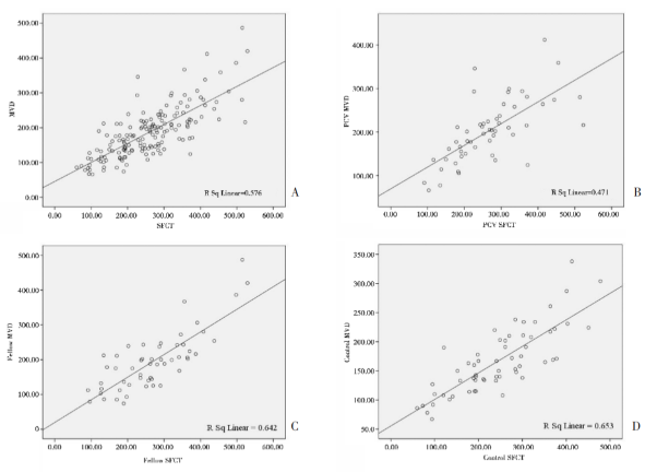 A. Overall corelation analysis between SFCT and MVD; B. Correlation analysis of SFCT and MVD in the PCV affected eyes; C. Correlation analysis of SFCT and MVD in the fellow eyes of PCV patients; D. Correlation analysis of SFCT and MVD in the normal eyes of healthy subjects
No statistical difference was found among the three groups in terms of SFCT by single-factorial ANOVA (P=0.335; group A: 266.45±99.51 μm, group B: 269.57±105.10 μm and group C: 243.83±99.68 μm). Paired comparisons revealed no statistically significant differences (P=0.238 for groups A and C, P=0.875 for groups A and B and P=0.180 for groups B and C). Single-factorial ANOVA revealed a statistical difference in MVD among the three groups (P=0. 020) . Paired comparison showed that MVD significantly differed between groups A and C (P=0. 008) and between groups B and C (P=0. 038) , whereas no significant difference was noted between groups A and B (P=0. 576) , as indicated in Table 1.

Recent studies reported that the eyes affected with PCV can be classified into those with/without choroidal vascular hyperpermeability¹⁰. However, 12 month anti-VEGF treatment demonstrated that the best corrected visual acuity (BCVA) was significantly improved from 0.68 to 0.5 (logMAR) in the low perfusion group, whereas the BCVA was slightly enhanced from 0.79 to 0.74, hinting that perfusion sta- tus exerts a significant effect upon the prognosis of BCVA in PCV patients". Jirarattanasopa et al. proposed that the choroidal thickness of PCV patients is correlated with the polymorphism of 162V CFH gene12. In the present study, no signs of thickened SFCT was found,which is inconsistent with the findings of Chung⁴and Yang⁵, and may reflect the presence of different polymorphisms. Pichai Jirarattanasopa et al. found that ICGA angiography revealed both low and high choroidal perfusion in either typical wAMD or PCV patients¹², indicating that high choroidal perfusion is not specific to PCV or a prerequisite for the incidence of PCV. In certain PCV patients, choroidal vascular abnormality and hemangioma tissues were not caused by high perfusion, probably due to alternative factors such as inflammation and similar changes³. Our indings demonstrated that a variety of factors may cause PCV-like fundus lesions. Inflammation and oxidative stress probably play a role in this process,and may be factors that lead to the high incidence of PCV in the Asian male population.
For convenient observation, SFCT has been utilized as the primary index of choroidal thickness measurement. However, PCV is most likely to be found outside the macular fovea, accounting for approximately 63%14. Measuring subfoveal choroidal thickness alone cannot reflect the overall choroidal condition at the posterior pole of the fundus. Yang et al15. proposed the concept of "the largest choroidal vessel diameter,"which was defined as the largest diameter of choroidal hyporeflective lumina within the entire macular area. Yang et al. employed SD-OCT to measure choroidal MVD in PCV patients and found that the mean choroidal MVD of the PCV-affected eyes was (236±63) μm, significantly larger than (137±48) μm of the unaffected fellow eyes. In the current study,the mean choroidal MVD of the PCV-affected eyes was (202.55±72.45) μm,equally significantly larger compared with (166.45±56.18) μm of the healthy controls (P=0.008). The mean MVD of the patients'unaffected fellow eyes was thicker than that of the normal controls, whereas no difference was noted in the SFCT of the same group. MVD is a maximal value measured on a relatively wide range, and has a higher sensitivity for reflecting the status of choroidal perfusion compared with other indexes.
At present, SFCT and MVD measurement by SD-OCT should be aided by manual operation. In this study, the error could not be completely eliminated in spite of repeated measurements. In addition, a substantial number of PCV patients with subretinal hemorrhage were excluded from this clinical trial. Consequently,the patients enrolled in this study can not represent the entire population of PCV patients. This potentially explains why no statistical significance was found in SFCT between the two groups. Subsequent investigation of choroidal morphology and function in PCV patients is likely to contribute to revealing the underlying mechanism.
The results in the present study demonstrated that MVD was positively correlated with SFCT in both PCV patients and normal subjects. No statistical significance was found in SFCT between PCV affected and healthy eyes. The MVD was thicker in the PCV affected eyes than in unaffected normal eyes. Moreover, the MVD was equally thick in the unaffected fellow eyes and in the normal controls, hinting that choroidal high perfusion constantly affected bilateral eyes. The fellow eyes had a high risk of PCV, which should be closely followed up and properly treated. In addition, MVD is a highly sensitive index that reflects choroidal perfusion, and is worth further investigation.
Choroid vascular pathological dilatations may underlie the pathogenesis of PCV².Terasaki et al³. Obtained submacular tissue specimens from PCV patients intraoperatively and found dilated thin-walled vessels in Bruch's membrane with multiple lesions in polypoidal or cluster shapes. Indocyanine green angiography revealed the formation of high-perfusion angiopathy of the choroidal vasculature and polypoidal structure, further confirming the signs of PCV. At present, this is considered the gold standard for PCV diagnosis.
The advances in optic coherence tomography (OCT) have led to a deeper understanding of the underlying pathogenesis of PCV. The emergence of newgeneration spectral domain optical coherence tomography (SD-OCT) allows explicit observation of each layer structure of the retina and choroid4,5. In the present study, the alterations in choroidal vasculature were observed in PCV patients by using SD-OCT to measure the subfoveal choroidal thickness (SFCT) and choroidal maximal vesseldiameter (MVD) in affected and unaffected fellow eyes.
Materials and methods
Study subjects
In this cross-sectional study, 53 consecutive patients (53 eyes) diagnosed with active PCV between 2010 and 2013 were enrolled and another 60 age- and gender- matched healthy subjects (60 eyes) were enrolled as normal controls.The final diagnosis of active PCV was confirmed by two experienced physicians from the Department of Fundus Diseases. The third physician was required to validate the diagnosis when their opinions varied. All patients underwent slit-lamp examination, preset lens examination, SD-OCT, fundus fluorescein angiography (FFA), and ICGA. Diagnosis criteria of PCV:ICGA showed characteristic polypoidal lesions, accompanied with/without abnormal choroidal vessel network⁶. Exclu- sion criteria included:1. Those patients with bilateral PCV; 2. Those complicated with other ocular pathological changes, such as glaucoma, histoplasma capsulatum infection,history of intraocular surgery, ocular contusion,amblyopia,and media opacity, or alternative fundus pathological changes, such as diabetic retinopathy, uveitis, pathological myopia, proliferative retinal diseases, retinal atrophy,macular epiretinal membranes,or age-related macular degen eration; 3. Those with subretinal hemorrhage affecting ICGA and OCT; and 4. Those had undergone anti-VEGF, photodynamic therapy,intraocular injection of triamcinolone acetonide, or thermal laser therapy, etc.Measurement methods
All patients were subject to SD-OCT (Heidelberg, Germany) for enhanced depth scans7. OCT was performed to conduct 7 scans of the posterior polar region within a rectangular area of 5×30 degrees using 8.8 μm scanning segments through the macular fovea. Each OCT image was constructed from 100 overlapped images. Choroidal thickness was defined as the zonal area between the outer surface of the retinal pigment epithelium and the inner scleral surface.Maximal diameters of choroidal vessels were measured from OCT data obtained from vertical and horizontal sections (4500 μm×750 μm) under the center of the fovea. The maximal vessel diameter was selected in the large blood vessels,measured vertically to the Bruch's membrane. All measurement procedures were accomplished independently by two experienced physicians. The data consistency was analyzed by statistical methods. The average of two measurements was calculated when the inconsistency degree was≤10%. Repeated measurement was required when the inconsistency degree exceeded 10%.Statistical analysis
SPSS 16.0 software was employed for statistical analysis. Patient age between the case and control groups was compared by an independent sample t-test,and gender was compared by a chsquare test. Choroidal thickness and maximal vessel diameter among the affected and unaffected fellow eyes and normal subjects were statistically analyzed by paired t-test. Two groups comparisons among three groups (group A: affected eyes,group B: fellow eyes and group C: healthy eyes) were conducted by using LSD tests. The correlation between SFCT and MVD was evaluated by Pearson correlation analysis. P<0.05 was considered as statistically significant.Results
Fifty three patients (53 eyes) were enrolled in this clinical trial, including 18 males and 35 females, aged 63.2±8.0 years on average. Sixty healthy subjects comprised the control group including 28 males and 32 females,aged 65.5±8.7 years on average. No statistical difference was observed between case and control groups regarding gender (P=0.170) or age (P=0.151). Overall analysis revealed a linear correlation between MVD and SFCT (P<0. 001; r=0. 759).Among the three groups, MVD was correlated with SFCT in a linear pattern(P<0. 001, r=0. 686; P<0. 001, r=0. 801; P<0. 001, r=0.808). The correlation coefficient was slightly lower for group A than for groups B and C, as shown in Figure 1.
Figure 1
No statistical difference was found among the three groups in terms of SFCT by single-factorial ANOVA (P=0.335; group A: 266.45±99.51 μm, group B: 269.57±105.10 μm and group C: 243.83±99.68 μm). Paired comparisons revealed no statistically significant differences (P=0.238 for groups A and C, P=0.875 for groups A and B and P=0.180 for groups B and C). Single-factorial ANOVA revealed a statistical difference in MVD among the three groups (P=0. 020) . Paired comparison showed that MVD significantly differed between groups A and C (P=0. 008) and between groups B and C (P=0. 038) , whereas no significant difference was noted between groups A and B (P=0. 576) , as indicated in Table 1.
Table 1 Analysis between Subfoveal choroidal thickness (SFCT) and maximum vessel diameter (MVD) in each group
Discussion
PCV is described as a peculiar hemorrhagic and exudative disorder of the choroid, characterized by recurrent serous or hemorrhagic detachment of the subretinal and sub-retinal pigment epithelium1,8. Previous studies demonstrated that the SFCT of the PCV affected eyes was thickened compared with that in normal subjects. Chung et al⁴. reported the mean SFCT of affected eyes from 25 PCV patients (438± 88) μm was significantly greater than (373±112) μm of 14 fellow eyes and (225±53) μm of the normal controls. Yang et al⁵. equally confirmed that the average SFCT of 13 PCV affected eyes (338±107 μm) was significantly greater than the value (261±78 μm) in healthy subjects. However, these two trials were both conducted in Asian populations and the sample sizes were limited. A thicker SFCT has been demonstrated in PCV patients compared to wAMD⁹ . Nevertheless,the outcomes in this clinical trial revealed that although the mean SFCT was greater in the affected eye group than in the control group, no statistical difference was observed among the three groups in terms of mean SFCT (P=0. 238,P=0. 875 and P=0. 180) .Recent studies reported that the eyes affected with PCV can be classified into those with/without choroidal vascular hyperpermeability¹⁰. However, 12 month anti-VEGF treatment demonstrated that the best corrected visual acuity (BCVA) was significantly improved from 0.68 to 0.5 (logMAR) in the low perfusion group, whereas the BCVA was slightly enhanced from 0.79 to 0.74, hinting that perfusion sta- tus exerts a significant effect upon the prognosis of BCVA in PCV patients". Jirarattanasopa et al. proposed that the choroidal thickness of PCV patients is correlated with the polymorphism of 162V CFH gene12. In the present study, no signs of thickened SFCT was found,which is inconsistent with the findings of Chung⁴and Yang⁵, and may reflect the presence of different polymorphisms. Pichai Jirarattanasopa et al. found that ICGA angiography revealed both low and high choroidal perfusion in either typical wAMD or PCV patients¹², indicating that high choroidal perfusion is not specific to PCV or a prerequisite for the incidence of PCV. In certain PCV patients, choroidal vascular abnormality and hemangioma tissues were not caused by high perfusion, probably due to alternative factors such as inflammation and similar changes³. Our indings demonstrated that a variety of factors may cause PCV-like fundus lesions. Inflammation and oxidative stress probably play a role in this process,and may be factors that lead to the high incidence of PCV in the Asian male population.
For convenient observation, SFCT has been utilized as the primary index of choroidal thickness measurement. However, PCV is most likely to be found outside the macular fovea, accounting for approximately 63%14. Measuring subfoveal choroidal thickness alone cannot reflect the overall choroidal condition at the posterior pole of the fundus. Yang et al15. proposed the concept of "the largest choroidal vessel diameter,"which was defined as the largest diameter of choroidal hyporeflective lumina within the entire macular area. Yang et al. employed SD-OCT to measure choroidal MVD in PCV patients and found that the mean choroidal MVD of the PCV-affected eyes was (236±63) μm, significantly larger than (137±48) μm of the unaffected fellow eyes. In the current study,the mean choroidal MVD of the PCV-affected eyes was (202.55±72.45) μm,equally significantly larger compared with (166.45±56.18) μm of the healthy controls (P=0.008). The mean MVD of the patients'unaffected fellow eyes was thicker than that of the normal controls, whereas no difference was noted in the SFCT of the same group. MVD is a maximal value measured on a relatively wide range, and has a higher sensitivity for reflecting the status of choroidal perfusion compared with other indexes.
At present, SFCT and MVD measurement by SD-OCT should be aided by manual operation. In this study, the error could not be completely eliminated in spite of repeated measurements. In addition, a substantial number of PCV patients with subretinal hemorrhage were excluded from this clinical trial. Consequently,the patients enrolled in this study can not represent the entire population of PCV patients. This potentially explains why no statistical significance was found in SFCT between the two groups. Subsequent investigation of choroidal morphology and function in PCV patients is likely to contribute to revealing the underlying mechanism.
The results in the present study demonstrated that MVD was positively correlated with SFCT in both PCV patients and normal subjects. No statistical significance was found in SFCT between PCV affected and healthy eyes. The MVD was thicker in the PCV affected eyes than in unaffected normal eyes. Moreover, the MVD was equally thick in the unaffected fellow eyes and in the normal controls, hinting that choroidal high perfusion constantly affected bilateral eyes. The fellow eyes had a high risk of PCV, which should be closely followed up and properly treated. In addition, MVD is a highly sensitive index that reflects choroidal perfusion, and is worth further investigation.
基金
暂无基金信息
参考文献
1. Yannuzzi LA,Sorenson J,Spaide RF,et al. Idiopathic polypoidal choroidal vasculopathy (Ipcv) . 1990. Retina, 2012,32 Suppl 1:1-8.
2. Okubo A,Sameshima M,Uemura A,et al. Clinicopatho- logical correlation of polypoidal choroidal vasculopathy revealed by ultrastructural study. Br J Ophthalmol,2002, 86 (10) :1093-1098.
3. Terasaki H,Miyake Y,Suzuki T,et al. Polypoidal choroidal vasculopathy treated with macular transloca- tion:clinical pathological correlation. Br J Ophthalmol, 2002,86 (3) :321-327.
4. Chung SE,Kang SW,Lee JH,et al. Choroidal thickness in polypoidal choroidal vasculopathy and exudative age- related macular degeneration. Ophthalmology,2011,118 (5) :840-845.
5. Yang LH,Jonas JB,Wei WB. Optical coherence tomo- graphic enhanced depth imaging of polypoidal choroidal vasculopathy. Retina,2013,33 (8) :1584-1589.
6. lida T. Diagnostic criteria for polypoidal choroidal vascu- lopathy and their clinical significance. Nihon Ganka Gakkai Zasshi,2005,109 (7) :383-384.
7. Spaide RF,Koizumi H,Pozzoni MC. Enhanced depth imaging spectral-domain optical coherence tomography. Am J Ophthalmol,2008,146 (4) :496-500.
8. Maruko I,lida T,Saito M,et al. Clinical Characteristics of exudative age-related macular degeneration in japanese patients. Am J Ophthalmol,2007,144 (1) :15-22.
9. H. Koizumi,T. Yamagishi,T. Yamazaki,et al. Subfoveal Choroidal Thickness in Typical Age-Related Macular De- generation and Polypoidal Choroidal Vasculopathy. Grae- fes Arch Clin Exp Ophthalmol. 2011. 249. 1123-1128.
10. Maruko I,lida T,Sugano Y,et al. Subfoveal retinal and choroidal thickness after verteporfin photodynamic thera- py for polypoidal choroidal vasculopathy. Am J Ophthalmol,2011,151 (4) :594-603 el.
11. Nakata I,Yamashiro K,Kawaguchi T,et al. Association between the cholesteryl ester transfer protein gene and polypoidal choroidal vasculopathy. Investigative Ophthal- mology &Visual Science,2013,54 (9) :6068-6073.
12. Jirarattanasopa P,Ooto S,Nakata I,et al. Choroidal thickness,vascular hyperpermeability,and complement factor h in age-related macular degeneration and poly- poidal choroidal vasculopathy. Invest Ophthalmol Vis Sci, 2012,53 (7) :3663-3672.
13. MacCUMBER MW,Dastgheib K,Bressler NM,et al. Clinicopathologic correlation of the multiple recurrent serosanguineous retinal pigment epithelial detachments syndrome. Retina,1994,14 (2) :143-152.
14. Cackett P,Htoon H,Wong D,et al. Haemorrhagic pig- ment epithelial detachment as a predictive feature of polypoidal choroidal vasculopathy in a chinese popula- tion. Eye (Lond) ,2010,24 (5) :789-792.
相关文章
孟丽慧;于仟仪;杨景元;陈有信,Application of anterior scleral thickness measurement in different ocular conditions using anterior segment optical coherence tomography: a systemic review甘雨虹;文峰;周孝来,A man with asymmetric bilateral choroidal melanocytosis: a case report and literature review丘蔚晴;张夏茵;赵兰琴;刘冬;温静怡;陈霞;王倩;李慧群;陈彦茹;韩彤;叶增纯;吴倩妮;林铎儒;肖辉;彭晖;林浩添,Retinal neurovascular characteristics for the diagnosis and staging of nondiabetic chronic kidney disease: a diagnostic study