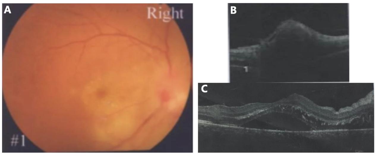
患者为男性,47岁,因“右眼无痛性视力下降3周”于2019年8月9日至中山大学中山眼科中心眼免疫与葡萄膜炎科就诊,否认眼红、头痛、发热等伴随症状。既往史、个人史、家族史无特殊。起病1周时患者于当地医院初诊时右眼检查资料见图1。
转诊至我科就诊时专科体格检查:最佳矫正视力(best corrected visual acuity, BCVA) OD 0.32 OS 1.0,眼压正常,双眼眼前节未见明显异常,右眼玻璃体细胞(+),视盘水肿,边界欠清,黄斑区大量黄白色星芒状渗出,左眼眼底未见明显异常(图2)。
Figure 1 Fundus photography and OCT findings of case 1 at initial presentation (1 week after symptom onset)
(A)右眼视盘水肿,边界不清,后极部视网膜水肿、黄色渗出;(B~C)OCT可见视盘及黄斑区神经上皮层脱离。
(A) Right eye showed optic disc edema with blurred margins, retinal edema, and yellow exudates in the posterior pole. (B-C) OCT revealed
neurosensory detachment at the optic disc and macular.
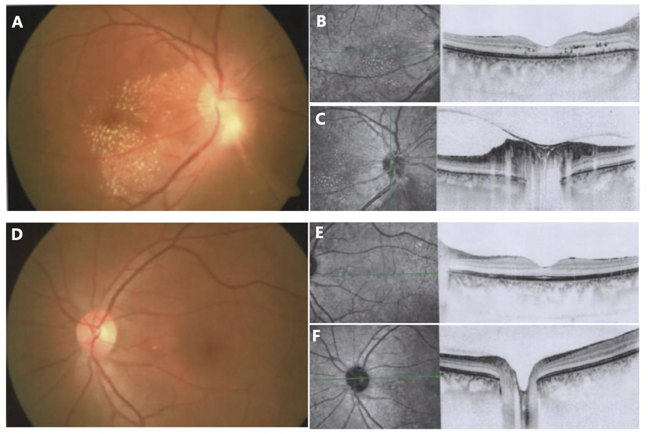
图2 例1患者转至我科就诊时(起病3周)的眼底表现及OCT改变
Figure 2 Fundus and OCT features of case 1 at referral visit (3 weeks after onset)
(A~C)右眼视盘水肿,边界欠清,黄斑区大量黄白色星芒状渗出;OCT示右眼视网膜外丛状层、外核层多个点团状高反射
信号病灶,椭圆体带连续性欠佳,视盘水肿。(D~F)左眼眼底及OCT未见明显异常。
(A-C) Right eye demonstrated optic disc edema with ill-defined margins and abundant yellowish-white stellate exudates in the macula. OCT
showed multiple punctate hyperreflective foci in the outer plexiform and outer nuclear layers, disrupted ellipsoid zone continuity, and optic disc
edema. (D-F) Left eye exhibited unremarkable fundus and OCT findings.
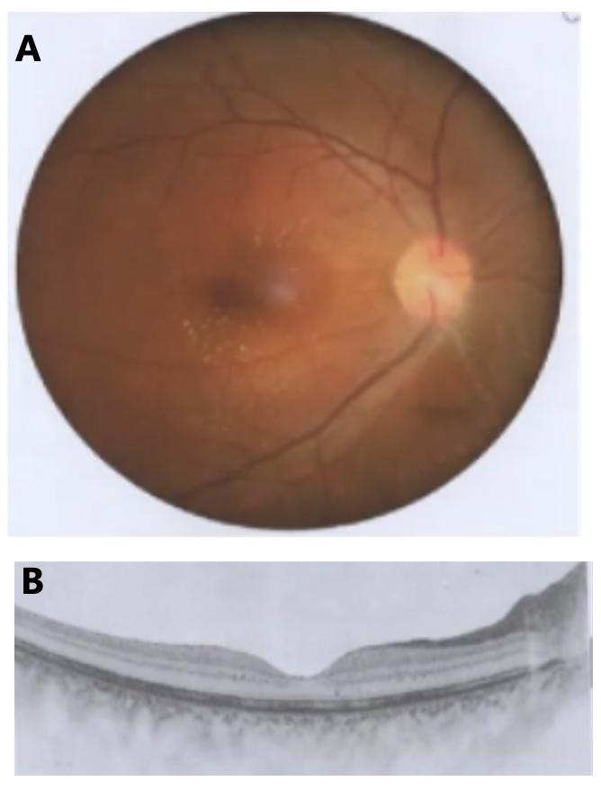
图3 右眼治疗3月后眼底表现及OCT改变
Figure 3 Post-treatment fundus and OCT changes in the right
eye (3 months)
A:右眼视盘水肿消退,边界清,黄斑区星芒状渗出大部份吸收;B:OCT示黄斑区外从状层、外核层散在少量点状高反射信号灶。
(A) Resolution of optic disc edema with sharp margins and near complete absorption of macular stellate exudates. (B) OCT revealed
sparse punctate hyperreflective foci in the outer plexiform and outer
nuclear layers of the macula.
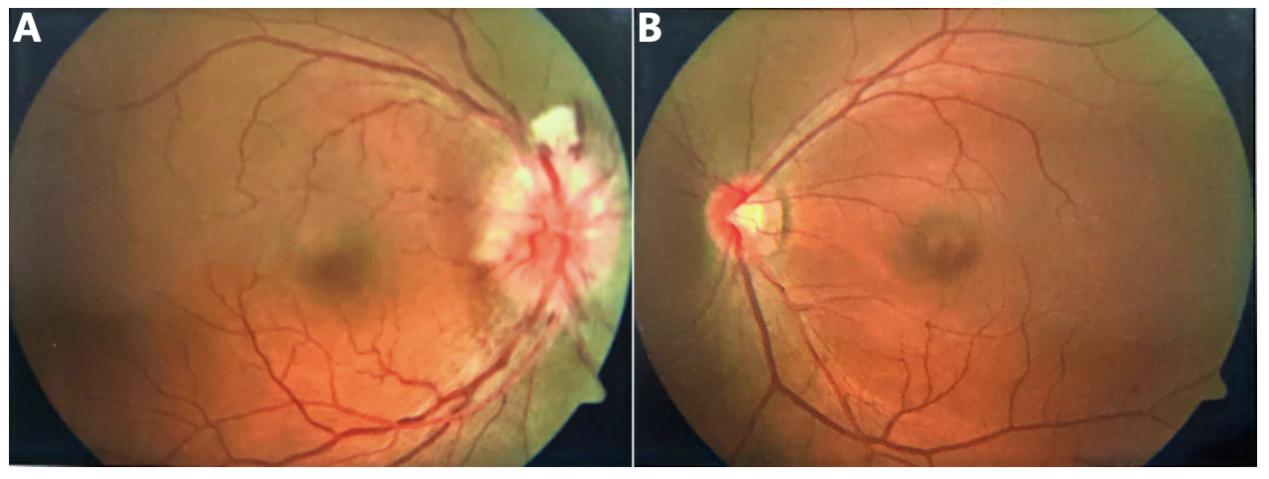
图4 例2患者双眼起病时眼底表现
Figure 4 Baseline fundus features of case 2
(A)右眼视盘充血水肿,边界不清,视盘上半表面线片状出血,片状棉绒斑,后极部血管稍迂曲扩张,黄斑中心凹反光尚可
见;(B)左眼眼底未见明显异常。
(A) Right eye showed hyperemic optic disc edema with blurred margins, linear hemorrhages on the superior disc surface, cotton wool spots,
mildly tortuous retinal vessels in the posterior pole, and preserved foveal reflex. (B) Left eye with normal fundus.
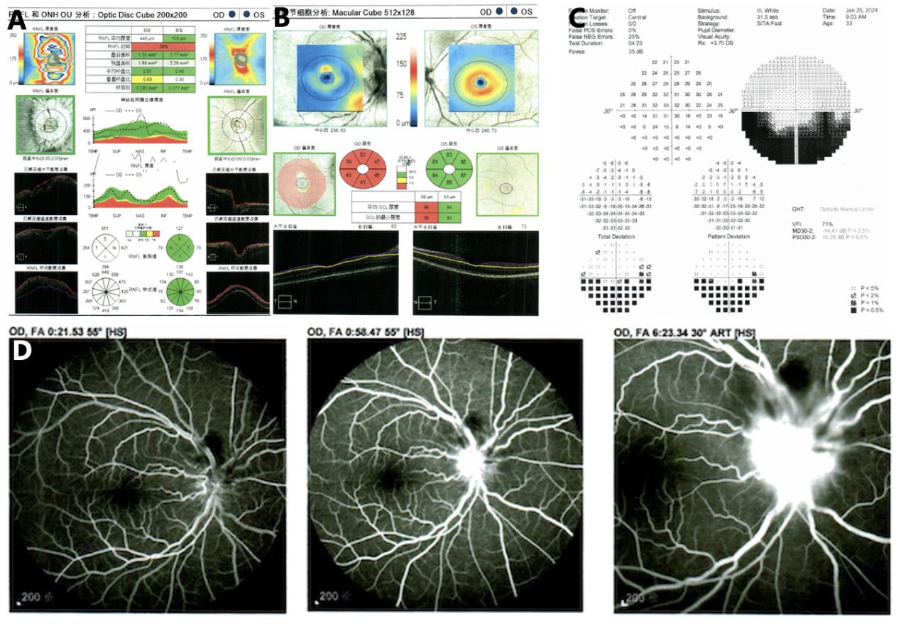
图5 例2患者起病时右眼OCT、视野及FFA改变
Figure 5 Baseline OCT, visual field, and FFA findings of case 2 (right eye)
(A)右眼视盘RNFL肿胀;(B)右眼弥漫性神经节细胞萎缩;(C)右眼视野呈现弓形缺损;(D)右眼视盘表面毛细血管迂曲扩张
伴显著荧光素渗漏,晚期视盘强荧光,边界不清,视盘周围血管迂曲,视盘上方不规则片状遮蔽荧光(渗出及出血)。
(A) Swelling of the peripapillary retinal nerve fiber layer (RNFL). (B) Diffuse ganglion cell layer atrophy. (C) Visual field demonstrating arcuate
defect. (D) FFA showed tortuous and dilated peripapillary capillaries with significant dye leakage, late-phase disc hyperfluorescence with
blurred margins, vascular tortuosity, and an irregular hypofluorescent area superior to the disc (corresponding to exudates/hemorrhage).
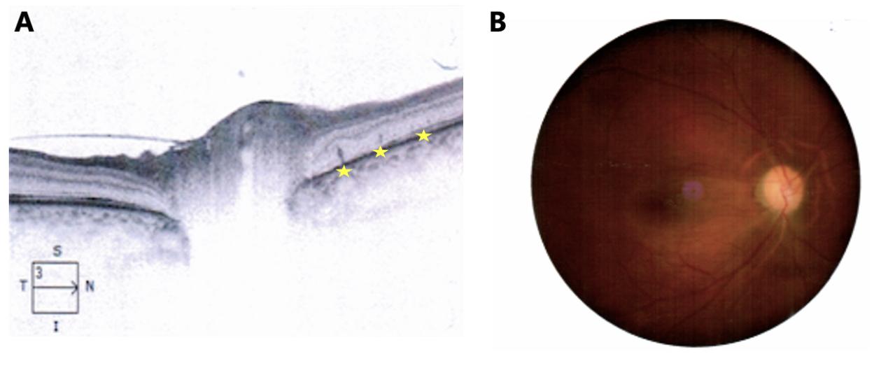
图6 例2患者治疗后右眼OCT及眼底表现
Figure 6 Post-treatment changes in case 2 (right eye)
(A)治疗1个月后,右眼视盘周围视网膜外层可见数个钉状突起高反射病灶(黄色星形指示);(B)驱梅治疗3个月后,右眼视盘
水肿消退,边界尚清,色泽苍白。
(A) OCT at 1-month follow-up: Focal spicule-like hyperreflective lesions in the peripapillary outer retina (yellow asterisks). (B) Fundus at 3
months after anti-syphilitic treatment: resolved disc edema with relatively distinct margins and pallor, indicating optic atrophy.
1. 国家自然科学基金(82301259;U22A20308);广州市科技计划项目(SL2023A04J00243,
SL2024A04J00243,SL2024A03J00513)。This work was supported by The National Natural Science Foundation of China
(82301259, U22A20308). Guangzhou Science and Technology Plan Project (SL2023A04J00243, SL2024A04J00243,
SL2024A03J00513).
2. Chi SL, Stinnett S, Eggenberger E, et al. Clinical characteristics in 53 patients with cat scratch optic neuropathy[J]. Ophthalmology, 2012, 119(1): 183-187. DOI: 10.1016/j.ophtha.2011.06.042.
3. Furtado JM, Simões M, Vasconcelos-Santos D, et al. Ocular syphilis[J]. Surv Ophthalmol, 2022, 67(2): 440-462. DOI: 10.1016/j.survophthal.2021.06.003.
4. Alhawsawi AA, Aljahdali A, Magharbil E, et al. The clinical spectrum and outcomes of ocular syphilis in Saudi Arabia: the emergence of a uveitic masquerader[J]. J Epidemiol Glob Health, 2025, 15(1): 31. DOI: 10.1007/s44197-025-00374-1.
5. Oska N, Saad M, Tokko H. Hypertensive disc edema or ocular syphilis? a case report of the great masquerader[J]. Case Rep Ophthalmol, 2025, 16(1): 346-352. DOI: 10.1159/000545491.
8. Perkins BA, Swaminathan B, Jackson LA, et al. Case 22-1992--pathogenesis of cat scratch disease[J]. N Engl J Med, 1992, 327(22): 1599-601. DOI: 10.1056/NEJM199211263272215.
9. Dehio C. Molecular and cellular basis of Bartonella pathogenesis[J]. Annu Rev Microbiol, 2004, 58: 365-390. DOI: 10.1146/annurev.micro.58.030603.123700.
10. Habot-Wilner Z, Trivizki O, Goldstein M, et al. Cat-scratch disease: ocular manifestations and treatment outcome[J]. Acta Ophthalmol, 2018, 96(4): e524-e532. DOI: 10.1111/aos.13684.
11. Abdelhakim A, Rasool N. Neuroretinitis: a review. Curr Opin Ophthalmol, 2018, 29(6): 514-519. DOI: 10.1097/ICU.0000000000000527.
12. Reed JB, Scales DK, Wong MT, et al. Bartonella henselae neuroretinitis in cat scratch disease[J]. Ophthalmology, 1998, 105(3): 459-466. DOI: 10.1016/s0161-6420(98)93028-7.
13. Ormerod L. Retinal and choroidal manifestations of cat-scratch disease[J]. Ophthalmology, 1998, 105(6): 1024-1031. DOI: 10.1016/s0161-6420(98)96003-1.
14. Jurke A, Bannert N, Brehm K, et al. Serological survey of Bartonella spp., Borrelia burgdorferi, Brucella spp., Coxiella burnetii, Francisella tularensis, Leptospira spp., Echinococcus, Hanta-, TBE- and XMR-virus infection in employees of two forestry enterprises in North Rhine–Westphalia, Germany, 2011–2013[J]. Int J Med Microbiol, 2015, 305(7): 652-662. DOI: 10.1016/j.ijmm.2015.08.015.
17. Stepanić M, Duvnjak S, Reil I, et al. Epidemiology of Bartonella henselae infection in pet and stray cats in Croatia with risk factors analysis[J]. Parasites Vectors, 2024, 17(1): 48. DOI: 10.1186/s13071-024-06117-8.
18. Fabbi M, Vicari N, Tranquillo M, et al. Prevalence of Bartonella henselae in stray and domestic cats in different Italian areas: evaluation of the potential risk of transmission of Bartonella to humans[J]. Parassitologia, 2004, 46(1-2):127-129.
19. Chomel BB, Boulouis HJ, Petersen H, et al. Prevalence of Bartonella infection in domestic catsin Denmark[J]. Vet Res, 2002, 33(2): 205-213. DOI: 10.1051/vetres: 2002008.
20. Kabanovski A, Donaldson L, Jeeva-Patel T, et al. Optic disc edema in syphilis[J]. J Neuro Ophthalmol, 2022, 42(1): e173-e180. DOI: 10.1097/wno.0000000000001302.
'%20fill='white'%20fill-opacity='0.01'/%3e%3cmask%20id='mask0_3477_29692'%20style='mask-type:luminance'%20maskUnits='userSpaceOnUse'%20x='0'%20y='0'%20width='16'%20height='16'%3e%3crect%20id='&%23232;&%23146;&%23153;&%23231;&%23137;&%23136;_2'%20x='16'%20width='16'%20height='16'%20transform='rotate(90%2016%200)'%20fill='white'/%3e%3c/mask%3e%3cg%20mask='url(%23mask0_3477_29692)'%3e%3cpath%20id='&%23232;&%23183;&%23175;&%23229;&%23190;&%23132;'%20d='M14%205L8%2011L2%205'%20stroke='%23333333'%20stroke-width='1.5'%20stroke-linecap='round'%20stroke-linejoin='round'/%3e%3c/g%3e%3c/g%3e%3c/svg%3e)