





Anterior segment optical coherence topography and ultrasound biomicroscopy (UBM) are
two main anterior segment scanning and imaging tools for the cornea,
anterior chamber, chamber angle, iris, partial lens, and anterior
segment vitreous. previous studies revealed that both AS-OCT and UBM
could quantitatively analyze anterior segment structures such as central
corneal thickness, anterior chamber depth, chamber angle, etc., with a
relatively sound reproducibility and consistency1,2. However,
few studies have analyzed the reproducibility and consistency of AS-OCT
and UBM in measuring iris parameters in glaucoma eyes. Iris morphology
is one of the common risk factors for chamber angle closure and angle
closure glaucoma. Peripheral iris thickness (PIT) and iris curvature
(IC) are regarded as vital parameters in identifying the pathogenesis of
angle closure glaucoma3. In this study, AS-OCT and UBM were
utilized to perform anterior segment scanning on patients with primary
angle closure glaucoma and their reproducibility and consistency in
measuring iris parameters were compared, with the aim of evaluating
their application in clinical research on glaucoma.
subjects and methods
study subjects
A total of 22 patients (38 eyes) with angle closure glaucoma admitted as outpatients in zhongshan Ophthalmic Center between February and May 20l2 were enrolled in this study. The patients were 4 males and 18 females, aged 57.9±8.4 (40 to 77) years. Of the 38 eyes, 5 had acute angle closure glaucoma, 23 had chronic angle closure glaucoma, and 10 fellow eyes had acute angle closure glaucoma.
Inclusion criteria: ① aged ≥40 years; ② acute or chronic IOP eleVation > 21mmHg; ③ angle closure or presenting angle closure when eleVated IOP was detected by gonioscopy; patients with acute angle closure glaucoma had at least two of the following symptoms (headache, ophthalmalgia, even nausea, vomiting, intermittent visual acuity decrease and iridization) and at least three of the following signs (conjunctival hyperemia, corneal epithelial edema, pupils mid-dilated and fixation, shallow anterior chamber, angle closure and glaucomatous fleck); ④ fellow eyes characterized as having a shallow anterior chamber and narrow chamber angle. Exclusion criteria: ① those undergoing antiglaucoma surgery or laser peripheral iridectomy; ② those presenting with uncontrollable IOP at the onset of acute glaucoma; ③ those with corrected Visual acuity <0.3 and unable to cooperate with fixed Visual field examination; ④ those with complicated ocular trauma or corneal lesions. The administration of miotic and other medicines capable of altering anterior segment structure was discontinued at 1 week before scanning. IOP was maintained below 25 mmHg.
Inclusion criteria: ① aged ≥40 years; ② acute or chronic IOP eleVation > 21mmHg; ③ angle closure or presenting angle closure when eleVated IOP was detected by gonioscopy; patients with acute angle closure glaucoma had at least two of the following symptoms (headache, ophthalmalgia, even nausea, vomiting, intermittent visual acuity decrease and iridization) and at least three of the following signs (conjunctival hyperemia, corneal epithelial edema, pupils mid-dilated and fixation, shallow anterior chamber, angle closure and glaucomatous fleck); ④ fellow eyes characterized as having a shallow anterior chamber and narrow chamber angle. Exclusion criteria: ① those undergoing antiglaucoma surgery or laser peripheral iridectomy; ② those presenting with uncontrollable IOP at the onset of acute glaucoma; ③ those with corrected Visual acuity <0.3 and unable to cooperate with fixed Visual field examination; ④ those with complicated ocular trauma or corneal lesions. The administration of miotic and other medicines capable of altering anterior segment structure was discontinued at 1 week before scanning. IOP was maintained below 25 mmHg.
Anterior segment scanning
All subjects underwent anterior segment scans by AS-OCT (visante OCT, Carl Zeiss Co Ltd. )4 and UBM (SW-3200L, Tianjin Suowei Electronic Technology Co., Ltd. )8 by two clinicians in a room with an illumination of 70~100Lux
(luminometer mode: TES-l339).One horizontal scanning image was stored
for measurement.
Iris parameter measurement
Iris parameters were assessed in the image
obtained by AS-OCT and UBM by one single clinician (C.D. ). The
clinicians were blinded to the patients/basic information and to the
results of the other scanning tool. Each image was measured twice using
the caliper in the software at an interVal of two weeks.The detailed
data of iris the parameters are shown in Figure 1.
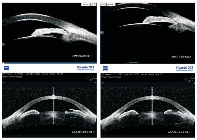
Peripheral iris thickness (PIT): the distance of two intersection points of the anterior and posterior iris surfaces by a line, about 500 μm from iris root, vertically passing through posterior surface of iris4. Iris convexity (IC): the maximum distance from the posterior surface of the iris to the line between pupillary margin and iris root, forward bowing as a positive value and backward dellen as negative5.
Figure 1 Iris parameters measured by AS-OCT and UBM
Peripheral iris thickness (PIT): the distance of two intersection points of the anterior and posterior iris surfaces by a line, about 500 μm from iris root, vertically passing through posterior surface of iris4. Iris convexity (IC): the maximum distance from the posterior surface of the iris to the line between pupillary margin and iris root, forward bowing as a positive value and backward dellen as negative5.
Statistical analysis
SPSS 18.0 software was employed for data
input and statistical analysis. For each type of scan, the
reproducibility of two measurements was expressed as the ICC of a
one-way random effects model, the data of two measurements were analyzed
in scatter diagram, and a pearson correlation efficient was calculated.
The first measurements of iris parameters by AS-OCT and UBM were
statistically compared by a paired t-test. The consistency between
AS-OCT and UBM was analyzed in a Bland-Altman scatter diagram. P
<0.05 was considered as statistically significant.
Results
Reproducibility of AS-OCT and UBM in quanti- tative analysis of iris parameters
The
ICC of PIT and IC, measured twice by AS- OCT, were 0.629 and 0.859,
respectively, and were 0.892 and 0.936 by UBM. Thus, UBM was superior to
AS-OCT in measurement reproducibility (Table 1). The correlation of
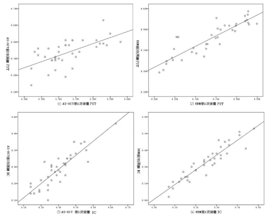
AS-OCT and UBM regarding repeatedly measuring iris parameters is shown
in Figure 2.
Table 1 Reproducibility of quantitative measurements of irisparameters by AS-OCT and UBM
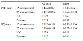
Figure 2 Correlation of repeated measurements of iris parameters by AS-OCT and UBM
consistency of AS-OCT and UBM in quantitative measurement of iris parameters
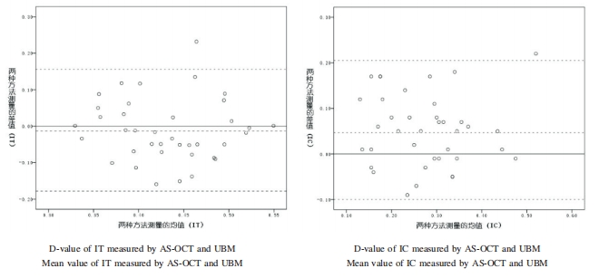
When measured by AS-OCT, the mean PIT of
38 eyes was 0.426 ±0.068 mm, which was 0.011 mm smaller than 0.438
±0.071 mm measured by ubM (t=一0.823,P=0.331), with the 95% LoA
(一0.178~0.156 mm) as 36.1~41.2% of the mean. The mean IC measurement was
0.302 ±0.104 mm by AS-OCT, which was significantly higher compared with
0.250±0.106 mm by ubM (t=4.lS7,P=0.017), with the 95% LoA (一0.100~0.206
mm) as 36.2~74.6% of the mean. (Table 2 and Figure 3)
Table 2 Consistency of quantitative measurements of iris parameters by A5-OCT and uBM

Figure 3 bland-Altman plot of iris parameters measured by AS-OCT and ubM
D-value of IT measured by AS-OCT and UBM
D-value of IC measured by AS-OCT and UBM
Mean value of IT measured by AS-OCT and ubM Mean value of IC measured by AS-OCT and ubM Figure 3 bland-Altman plot of iris parameters mea- sured by AS-OCT and ubM
Discussion
UBM with an ultrasonic frequency of 50MHz has adequate tissue penetration and yields images of anterior segment structures with an axial resolution as high as 50 μm. Pavlin et al4 reported that UBM gave good reproducibility in terms of quantitative measurement of anterior segment structures6,7. AS-OCT used a coherent optical signal acquisition and processing method at a wavelength of 1310 nm, and was performed by non-contact scanning in a sitting position. The axial resolution achieved was 18 μm. Due to the absorption by anterior segment tissues, such as the iris and ciliary body, the infrared laser was unable to penetrate into deep tissues so the images of the ciliary body or lens zonule were poor. The reproducibility in measuring chamber angle and central anterior chamber depth was confirmed as higher for AS-OCT than for UBM[2,8]. However, few studies have compared the application of AS-OCT and UBM regarding quantitative assessment of iris morphology.
The iris is a vital anterior segment tissue and plays an essential role in the pathogenesis of pupillary block and chamber angle closure in angle closure glaucoma. Iris parameters mainly include PIT and IC, and previous studies indicated that PIT and IC are independent factors associated with chamber angle narrowing9. A relatively thick iris is a risk factor for chamber angle closure10 and an important factor affecting the efficacy of laser peripheral iridectomy11. In the current study, the reproducibility and consistency of UBM and AS-OCT for quantitative measurements of iris thickness and iris curvature were statistically compared to evaluate the efficiency of these two tools in angle closure glaucoma research. The reliability and differences in the study results were also compared.
The reproducibility in measuring iris thickness and curvature was higher for UBM (ICC: 0.892 and 0.936) than for AS-OCT (ICC: 0.629 and 0.859). In addition, AS-OCT was not applicable for clinical practice due to its low reproducibility (<0.7) in measuring iris thickness (Table l and Figure 2). The difference in the reproducibility between AS-OCT and UBM may result from differing scanning principles between AS-OCT and UBM. AS-OCT emits infrared laser energy, but had relatively inadequate penetration across anterior segment tissues, especially the pigment layer of the iris and the ciliary body. The reference point of the iris root was not explicitly presented, making it difficult to locate. The UBM imaging produced full-thickness images of the ciliary body and the iris; the echo of the intersection point of the iris pigment epithelium and the ciliary process was complete and clear and the reference point of the iris root was easily located (Figure 1).
In addition, the measurement of mean PIC did not differ with AS-OCT and UBM (p=0.331); the 95% LoA(-0.178~0.156 mm) was still 36.1~41.2% of the mean. The IC measurement was smaller by UBM than by AS-OCT, with the 95% LoA(-0.100 ~ 0.206 mm) as 36.2~74.6% of the mean. The UBM test was performed with the subject in a supine position and eye position was fixed by gazing at a finger. In contrast, the AS-OCT was conducted from a sitting position and the eye position was fixed by gazing at an indicator light inside the machine. Gravity and accommodation, which could affect the iris morphology, were factors leading to different outcomes between UBM and AS-OCT. Another vital factor was the different scanning mode between UBM and AS-OCT. AS-OCT was performed at the primary posi- tion of eye. Images were captured at the site of the corneal central reflection band12 to maintain scanning deviation from corneal vertex within 0.8 mm and to avoid image distortion. During UBM scanning, the patients were told to constantly rotate their eyes to allow the probe vertical to the chamber angle tissues to acquire explicit images and relatively strong echo signaling. The anterior iris surface was also irregular due to iris texture, resulting in low reproducibility of the measured iris thickness. Accurate measurement of the iris area could reduce the influence of irregular iris morphology on the iris thickness measurement13.
In the current investigation, we compared the reproducibility and consistency between UBM and AS-OCT in measuring iris parameters and found a significant difference. The reproducibility was higher for UBM than for AS-OCT. Due to irregular iris morphology and different measurement procedures, both AS-OCT and UBM had a certain degree of variation. This degree of variation and discrepancy between UBM and AS-OCT should be taken into account when analyzing iris morphology in clinical practice to determine a reasonable sample quantity and equivalence of obtained results. Adopting area parameters and automatic analysis software will probably contribute to enhancing the accuracy and reproducibility of iris morphology measurement. Studies with larger sample sizes are required to further validate the outcomes.
In the current investigation, we compared the reproducibility and consistency between UBM and AS-OCT in measuring iris parameters and found a significant difference. The reproducibility was higher for UBM than for AS-OCT. Due to irregular iris morphology and different measurement procedures, both AS-OCT and UBM had a certain degree of variation. This degree of variation and discrepancy between UBM and AS-OCT should be taken into account when analyzing iris morphology in clinical practice to determine a reasonable sample quantity and equivalence of obtained results. Adopting area parameters and automatic analysis software will probably contribute to enhancing the accuracy and reproducibility of iris morphology measurement. Studies with larger sample sizes are required to further validate the outcomes.