





Introduction
Glaucoma is the leading cause of irreversible blindness worldwide1. Although primary angle-closure glaucoma (PACG) accounts for approximately 25% of all cases of this disease, it comprises 50%of bilateral blindness due to glaucoma2. PACG is more common in Asians than Whites3. In China, this form is a major cause of bilateral glaucoma blindness, accounting for 91% of cases4.Laser peripheral iridotomy (LPI) is a common initial treatment for PACG and has been shown to be effective in treating eyes with occludable anterior chamber angles related to pupillary block mechanism. In cases where the angles are still narrow after LPI, non-pupillary block mechanisms (such as plateau iris syndrome or lens-induced angle narrowing) often play a role.
Plateau iris configuration is characterized by anterior displacement of the ciliary processes, pushing the peripheral iris forward, typically causing narrow angles or angle closure5, 6. On slit-lamp examination, a flat iris plane and relatively normal central anterior chamber depth are key features of this condition6. Plateau iris syndrome refers to angle closure in the presence of plateau iris configuration and a patent LPI.6 Recent studies from Asia have demonstrated that plateau iris is present in about 30%of narrow angle eyes after LPI7, 8. The Liwan eye study found plateau iris configuration in 59%of narrow angle eyes in at least l quadrant after LPI9.
Ultrasound biomicroscopy (UBM) is an imaging device that provides objective, high-resolution images of the angle structures, including the ciliary body and anterior portion of the lens. This technology has been used to assess the mechanisms related to angle closure, including pupillary block and plateau iris.
Although the prevalence of plateau iris among Asians has been documented in a significant number of studies, there is relative paucity of data on the role of plateau iris in Caucasians. The aim of this study was to evaluate the prevalence of plateau iris syndrome diagnosed by UBM after LPI in Caucasians as compared to Asians in a U. S. clinic setting.
Materials and methods
This was a prospective, observational study of patients who underwent LPI in the glaucoma clinic at the University of California, San Francisco (UCSF) between January, 2008 and September, 2010. This study was approved by the Committee on Human Research at UCSF, and written informed consent was obtained.Clinical data that was gathered included visual acuity, intraocular pressure (IOP) by Goldmann applanation tonometry or pneumotonometry, slit lamp biomicroscopy, examination of the optic discs with a 90-diopter lens, gonioscopy, and central corneal thickness (CCT). Indentation gonioscopy was performed by a single glaucoma specialist using a Posner gonioprism (Model OPDSG, Ocular Instruments, Inc. , Bellevue, WA, USA)in a dark room setting, and the angles were graded using the Shaffer grading system. All patients underwent Humphrey visual field testing with a 30-2 pattern (Model HFA2, Zeiss Meditec, Dublin, CA, USA)and the Swedish Interactive Threshold Algorithm(SITA)program.
Patients were diagnosed as primary angle closure suspect (PACS), primary angle closure (PAC), or PACG. In this study, PACS was used as the category
for eyes that had an average Shaffer grade <2 among the 4 quadrants, and whose UBM showed iridotrabecular contact or slit angles in at least 2 quadrants without increased IOP, peripheral anterior synechiae (PAS), abnormal HVF, or glaucomatous optic neuropathy.
PAC is an eye which meets PACS criteria and has evidence of decreased functional trabecular meshwork due to contact with the peripheral iris such as PAS, increased IOP (IOP>21 mmHg), iris whorling, glaucomfleken, or excessive pigment deposition on the trabecular surface(but without criteria to be considered glaucoma)10.
PACG was defined as PAC with evidence of glaucoma that was diagnosed by using both structural damage of the optic nerve and functional damage indicated by characteristic visual field defects.
In this study, glaucomatous optic neuropathy was defined as:1) the vertical cup:disc ratio (VCDR) ≥0.7, CDR asymmetry ≥0.2, notching of the neuroretinal rim, or having a nerve fiber layer defect; and 2)the glaucoma hemifield test graded as “outside normal limits"and a cluster of three contiguous points at the 5%level on the pattern deviation plot, which was reproducible at least twice to assess glaucomatous visual field loss10. In patients unable to adequately perform a reliable field, a VCDR ≥0.9 was considered as glaucomatous optic neuropathy.
Patients with secondary narrow angle causes(such as iris cysts), post- trauma, or post surgery were excluded.
UBM (Model P45, Paradigm Medical, Inc. , Salt Lake City, UT, USA)was performed 4-6 weeks before and after LPI in a dark room using only one eye of each patient in the supine position with a 50 MHz probe. Radial scans of the nasal, inferior, temporal, and superior angles were obtained. In terms of image quality, the main goal was to record images in which the scleral spur (SS) and ciliary processes could be easily identified.
The images were randomized and interpreted qualitatively by a trained observer, and intra-observer reproducibility was assessed for the same observer.
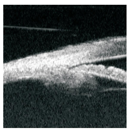
By UBM, plateau iris (Figures 1 and 2) was diagnosed in eyes that fulfilled all of the following criteria in at least two quadrants:
1. The ciliary process was directed anteriorly.
2. The ciliary sulcus was absent.
3. The central iris plane was flat.
Persistent appositional angles were diagnosed in eyes whose UBM showed iridotrabecular contact or slit angles after LPI in at least two quadrants.
Figure 1 Ultrasound biomicroscopy image from an eye with plateau iris showing iridotrabecular contact(A), anterior displacement of ciliary process (B), loss of the ciliary sulcus (C), and flat central iris plane(D). SS=Scleral spur.
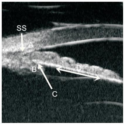
Figure 2 Ultrasound biomicroscopy image from an eye with plateau iris showing “slit” angle between peripheral iris and Cornea
Statistical analysis
Univariate comparisons of differences in categorical outcomes (the proportion of individuals with plateau iris) between ethnic classes were assessed using Fisher's exact test. Univariate comparisons of continuous outcomes between ethnic classes were conducted using the Wilcoxon rank-sum test. Multivariate analysis was conducted using logistic regression. Intra-observer reliability was assessed using the consistency ICC (intraclass correlation) coefficient. Statistical significance was assessed with an alpha of 0.05, two sided. All analyses were performed using R v. 2.12 for Macintosh (http://www.r-project.org ).Results
There were 63 subjects enrolled, each of whom received UBM imaging prior to and after their LPI. The images of eight subjects were excluded due to assessment of poor UBM quality in one or two quadrants by the reader. Fifty-five subjects were eligible for data analysis. There were 45 eyes with PACS, four eyes with PAC, and six eyes with PACG. Twenty-eight subjects(50. 09%) were Caucasian, and 27 subjects (49.1%)were Asians, com prised of 18 Chinese (66.7%), nine Filipino (33.3%), and one Vietnamese (3.7%). The mean age of the group was 683±10.9 years (range from 44 to 88 years); 31 of 55 subjects(56.4%)were females. The clinical characteristics of the two groups are shown in Table l. The overall mean IOP was 16. 8±4.0 mmHg (range from 9.5 to 28.5 mmHg), mean vertical CD ratio was 0.51±0.17(range from 0.25 to 0.90), and mean CCT was 538.5±34.6 μm (range from 470 to 610 μm).
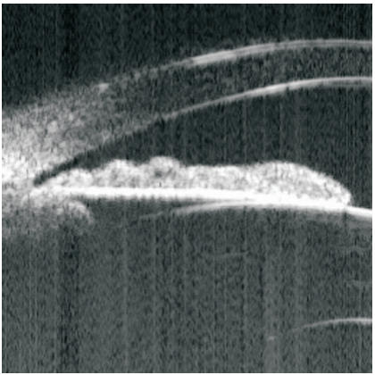
By UBM, persistent appositional angles were found in 28 of 55 subjects (50.9%), 15 of 28 Caucasians (53.6%), and thirteen of 27 Asians (48.1%) (no statistical difference between groups, P=0.79). We found persistent appositional angles with plateau iris in 18 of 55 subjects (32.7%), nine of 28 Cucasians (32.1%), and nine of 27 Asians (33.3%) (P>0.99). Plateau iris without appositional angles (Figure 3)were found in 15 of 55 subjects (27.3%), eight of 28 Caucasians (28.6%), and seven of 27 Asians (25.9%)(P>0.99) (Table 2).
The authors also analyzed the effect of ethnic classification on appositional angle adjusting for other variables individually:in particular, age, IOP, CDR, and CCT. There was no evidence of a difference in angle closure by ethnic category after these adjustments (P>0. 58). The authors also conducted multivariate logistic regression, adjusting for all these potential confounders, and continued to find no evidence of a difference by ethnic group (P=0.93). Inclusion of diagnostic category (PACS, PAC, or PACG) did not change these findings. The authors also modeled the presence or absence of plateau iris by ethnicity, and found no evidence of a univariate association (P=0.93), or when adjusting for age, gender, IOP, CDR, CCT individually (P>0.18), or all together (P=0.88). Inclusion of diagnostic category did not change these findings. Intraobserver reproducibility for diagnosing plateau iris with appositional angles for 64 UBM pictures was moderate (ICC=0.542).
Table 1 Clinical characteristics and biometric parameters of White and Asian subjects
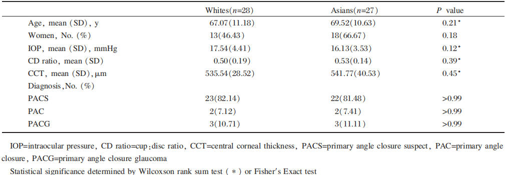
Table 2 Persistent appositional angles, plateau iris without appositional angles, and plateau iris with appositional angles

Figure 3 Ultrasound biomicroscopy image from an eye with features of plateau iris after laser peripheral iridotomy showing anterior ciliary process, absent ciliary sulcus, and flat central iris plane without iridotrabecular contact.
Discussion
In the present study, we found that residual narrow angles and plateau iris anatomy were common in both Asian and Caucasian populations seen within the same clinic. Pupillary block was once considered the overriding cause of narrow or closed angles. However, in the past several years, many studies have shown that non-pupillary block and multiple mechanisms are more common than previously thought. For example, in a review article in China, PACG due to multiple mechanisms (combination of pupillary block and iris crowding component, pupillary block and anteriorly positioned ciliary body, or all three together) was 54.8% of cases, followed by pupillary block alone (38.1%), then non-pupillary block mechanisms (7.8%)11.
In a UBM study in Indian eyes with PACG, there was persistent appositional angles after LPI in 60% of eyes(using angle opening distance [500 microns from SS] ≤130 microns in at least one quadrant as the criterion)¹². In the Liwan eye study, appositional angles were found in 59% of PACS eyes after LPI (at least one quadrant appositional as the criterion). In the present study, persistent appositional angles after LPI in PACS, PAC and PACG eyes (using at least 2 quadrants)were found in 53.6% of Caucasian eyes and 48.1% of Asian eyes. The percentage of persistent narrow angles in the present study may be slightly smaller than those in previous studies be cause most of the present subjects were PACS, and criteria required two or more quadrants to be narrow or closed. Taken together, these studies suggest that, even with a patent LPI, patients of different ethnicity still have a substantial risk from other mechanisms to develop PAS or angle closure glaucoma in the long term.
Using our UBM criteria, we found plateau iris with appositional angles in 33.3%of Asian eyes. This prevalence was similar to that of a study from Singapore, which found plateau iris after LPI in 32.3% of Chinese-Singaporean eyes with PACS7 . Furthermore, a recent study of PACG eyes in Singapore and Thailand also found the prevalence of plateau iris in similar proportions, 32. 8%and 31.7%, respectively8 .
Among Caucasians, we found plateau iris with appositional angles in 32.1%of eyes, which was not statistically different from Asians. Stieger et al. reported plateau iris syndrome in relatively young Caucasians (<60 years of age)with recurrent angle closure symptoms to be 47%¹³, and Ritch et al. found that 52% of subjects aged 40 years and younger with angle closure had plateau iris syndrome14.
There have been no reports about the prevalence of plateau iris syndrome in an older Caucasian population; however, pupillary block was believed to be the predominant cause. For example, in Lowe's study on fellow eyes of Caucasian patients with a cute angle closure (AAC), surgical PI prevented AAC in all patients, and 88.5% (23 of 26 fellow eyes) showed no glaucoma five years after the treatment15. Playfair and Watson's study had similar findings16. In the present study, the authors found a high rate of plateau iris in Caucasians, perhaps because UBM could demonstrate a greater detail of angle anatomy compared with clinical examination.
The authors also found plateau iris in subjects whose angles became open after LPI:27.3% of the entire group, 28.6% of Caucasian eyes, and 25.9% of Asian eyes, which is slightly higher than the report from Singapore(23.4%)7. The study from India also reported a high rate of anteriorly-positioned ciliary processes with absent ciliary sulcus in open angle eyes after LPI (40. 9%)12. These results suggest that plateau iris is a possible contributing mechanism in such eyes, even though pupillary block may be the major mechanism. Furthermore, a recent study in Japan found that 19.2% of POAG eyes had plateau iris configuration17.
Although the authors did not find plateau iris prevalence to be different between Caucasians and Asians, there may be other anatomical factors that contribute to the higher risk of angle closure in Asians, such as thicker iris, more anterior iris insertion, shallower anterior chamber depth (ACD), shorter axial length, and thicker lens. In a recent study using anterior segment optical coherence tomography, Leung et al. found that Chinese eyes had smaller ACD, smaller anterior chamber width, and greater iris thickness compared with Caucasian eyes18. The role of plateau iris in Caucasians and in POAG eyes deserves further studies.
There are some limitations with the present study.
Obtaining images from four quadrants of each eye shows only a small proportion of the overall angle anatomy. Furthermore, qualitative interpretation of the angles can be very subjective and observer-dependent. We performed an intra-observer reproducibility study, which showed moderate correlation. Intra-observer reproducibility in past studies was also moderate7, 8. Lastly, the number of subjects in each group was relatively small and the population was derived from a university-based clinic population, which has potential referral bias.
In conclusion, the prevalence of persistent narrow angles and plateau iris, as assessed by UBM, was similar in the Caucasian and Asian eyes within the clinic population. These results suggest that post-LPI monitoring of the angle and mechanism of action in Caucasians is imperative. Although pupillary block is involved in the majority of cases, other mechanisms, including plateau iris, are often contributory.